What an OCT scan shows, and why we offer one to everyone
A three-dimensional picture of the back of your eye, taken in seconds, that shows layers a photograph cannot.
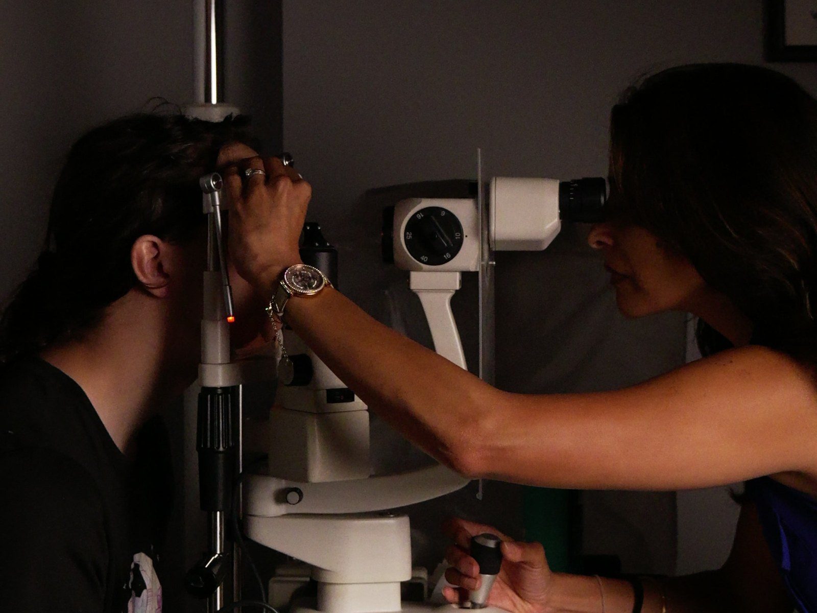
A cross-section, not a photograph
A retinal photograph shows the surface of the back of the eye. An OCT scan, which stands for optical coherence tomography, uses light to build a cross-section of the retina, layer by layer, in a few seconds and without anything touching your eye.
That cross-section is where the early signs of several conditions show first: changes at the macula, swelling, the thickness of the nerve fibre layer that matters in glaucoma.
Why we offer it to every patient
Because the most useful scan is the one taken before anything is wrong. Your first scan is a baseline. The next one is compared with it, and a change that would be invisible on its own becomes obvious side by side.
The scan is kept on your record. If you are referred to a hospital, the images go with you.
What it is not
An OCT scan is one part of an examination, not a substitute for it. It does not replace the tests of how you see, the look at the front of your eye, or the conversation about what you have noticed.